We are using bioglass, a bioactive ceramic that acts as a scaffold for the growing cells. The Image below shows the scaffold being initial shapped before it is seeded with cells. More images can be seen here.
ii) Kings College Workshop – Guys Hospital microbiology and imaging laboratory.
16th 17th May 2005.
I recently spent a couple of days wondering around the laboratories at Guys Hospital and met one of the PhD students who was kind enough to take me step by step through some of the processes involved in tissue engineering research. We began by looking at the raw materials used to produce the objects on which cell growth will eventually take place. I was shown a workshop not unlike the type I am usually acquainted with –drills, vices and metal cutting equipment are used to form sheets of titanium into small discs.
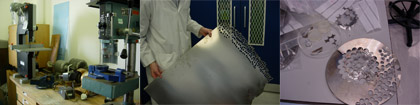
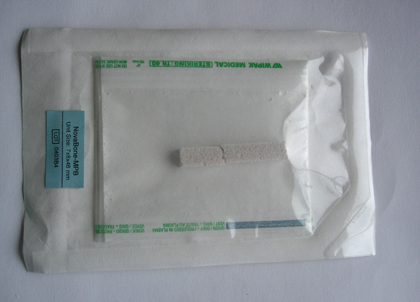
The titanium is cut to a specific size to fit the laboratory disposables and then treated with a coating of bioactive powder.
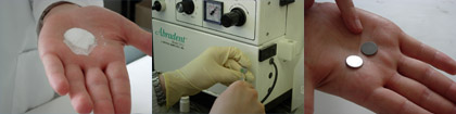
Titanium is widely used in for dental implants and is very biocompatible. The discs are sterilised to remove any harmful micro-organisms. This principle applies to the environment where tissue-culturing experiments - the process happens within a laminar flow hood which filters out any potential contamination from outside.
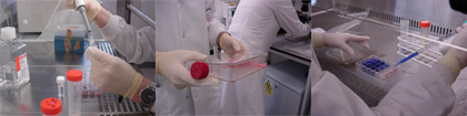
Keeping a laboratory space clean is the most important part of the whole process – the cultured cells are easily destroyed. The cells for tissue culture are housed temporarily in incubators, which maintain a constant body temperature of 37 degrees. The flasks, which contain primary cells, are filled with liquid nutrients, which promote cell growth.
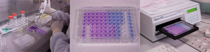
For the tissue engineer it is important to be able to quantify the cells within a sample. This is achieved by dying the cells with a blue stain, which differentiates the living cells from non-viable cells. The cells are continually supplied with fresh culture medium to keep them alive.
When the cells have been given adequate time to proliferate they are viewed using scanning electron microscopes and confocal microscopes. This will give an indication of how living cells react to particular types of bioglass.
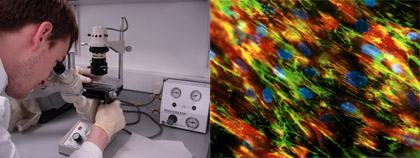
From using electron micro-graphs in my design work, I was interested in the process of how the images are achieved. The sample must be covered in a special oil so the microscope will give a clearer picture. The outcome of the cell culture experiments seemed fairly unpredictable. The first few titanium discs we looked at through a confocal microscope were very dark and I was unable to distinguish between the shapes of cells and background matter.
The fluorescent stain which had been previously added picked up on the live cells. The last disc revealed a spectacular image of the cells attached to the surface. I am not able to read the images as a scientist does, so my judgement of an image is an aesthetic one. Although to some extent so is theirs! As pieces of information they are unintelligible- I was given a quick lesson on how to interpret the image. A cell, which is ‘happy’ in its environment, will send out dendrites – like the roots of a plant spreading from the cell body onto the surface of the bioglass.
A camera is housed in the top of the microscope which allows movement over the surface of the sample to choose the clearest (and most appealing!) image.
We received the data and photographs of the samples I had prepared a few months ago. Unfortunately the samples had died due to contamination in the laboratory - they are currently being re-sterilized to begin the process again. Here are the images of the shaped bioglass in culture medium. The staining indicates that the cells liked the environment and had begun to grow.
iii) Tissue culture samples - report
The following images and text are taken from a report by Lucy D'Sliva who is working on the tissue culturing for the project. Click images to see a larger version:
Alamar blueTM assay post-incubation with test materials
The alamar blueTM (AB) is designed to quantitatively measure cell proliferation. This REDOX indicator responds to reduction or oxidation of the surrounding medium. It both fluoresces and changes colour in response to the chemical reduction of culture medium which results from cell growth and division. The greater the metabolic activity redox indicator changes from blue to pink.
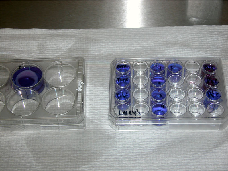
above: This plate shows samples 1-5 “stones” and sample 6 –“ring” immediately after addition of AB
The test samples are incubated in AB medium for a minimum of fours hours. As the cells grow and become metabolically active, innate metabolic activity results in a chemical reduction of AB.
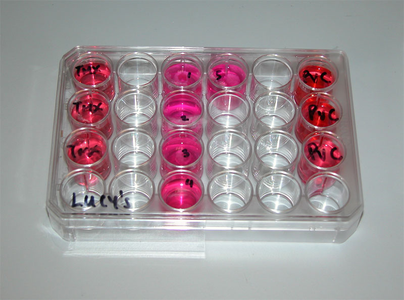
above: Continued growth maintains a reduced environment resulting in a change of colour from blue to pink.
TMX is Thermanox, a tissue culture control. PVC is a toxic control
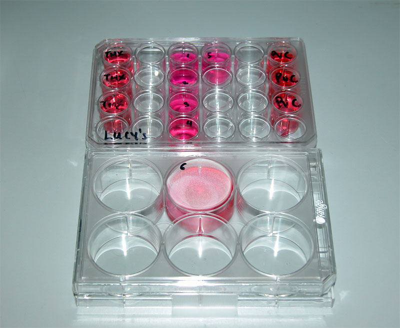
above: Samples 1-5 are the “stone” samples Sample 6 shows the intact ring in culture
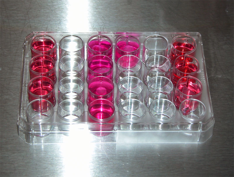
above: The bright pink colouration of the test samples in the middle rows indicates a high metabolic activity of proliferating cells