Donating cells through surgery
The cells used in culturing come from a range of sources. In Biojewellery we are using 2 sources.
Primary cells are provided from an anonymous donor, put in to storage with consent during a required medical operation. These differ from stem cells in that they are differentiated, which means they have descided what type of tissue they are, in our case bone tissue. We are using these cells for intial tests using sculpted bioglass.
Our second source of cells are from the couples who have volunteered to be a part of the project. All our couples need wisdom teeth taken out, and cells are taken from a fragment of bone, these chips are routinely removed as a part of the tooth extraction.
Hospitals need to follow strict guidelines when working with human cells and patient consent is a key part of this. It is important to us and our couples that when we design the rings we are using their cells.
i) Harvesting cells witn a biopsy
The initial plan was for our donor to undergo a biopsy, where cells are harvested from the femur. The image below shows this procedure. How were the cells used in this operation? Extracted cells were used in combination with a solid glass form of the biomaterial as a therapy for an injury to the bone sourrounding the donor's eye socket. The glass was shaped to fill the damaged area, and the bioactive properties of the material encouraged a strong bond between exisiting bone and the inserted glass. Cultured cells were added to help this bonding process.
In our case there is too much risk associated with the aneasthetic and surgery required for a biopsy. We are hoping to take a small bone chip from the donors jaw, when a wisdom tooth has been removed. It is common to remove chips of bone as a part of a wisdom tooth extraction. The challenge is keeping the cells that form this chip viable for cultuing, the sample must be kept in a sterile environment and frozen within a few hours.
ii) Images from wisdom teeth extraction
The images below are from an oral surgery theatre at Guys Hospital. These two operations were both wisdom tooth extractions, where general anaesthetic was used. Wisdom teeth are prone to decay and often cause overcrowding in the mouth which leads to other problems.
The eventual application for bioactive materials will be to grow tissue in vitro - or outside of the body - to repair parts of the body which have suffered disease or injury. An example of this would be the removal of a face and neck cancer, where bone tissue grown from the patients cells in vitro can then go back into the body. This avoids taking bone from the patients leg or arm, which creates a second operative site leading to more risk for the patient during the proceedure, more scaring after the operation.
This documentation of the wisdom teeth removal will be followed up later with images from a neck and face operation.
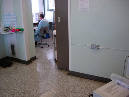
1. The theatre needs to be a hygenic safe envirnoment for the patient to avoid infection.
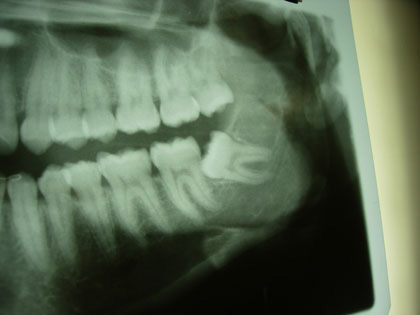
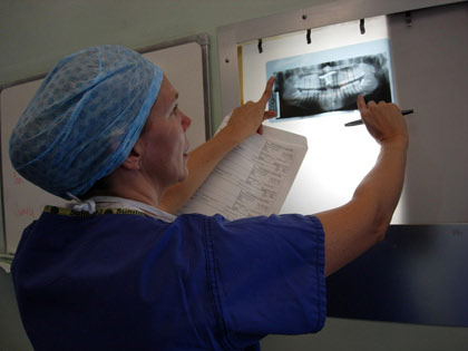
2. An x-ray of the first patient shows on of the problem teeth, it's growing at an angle putting pressure od the other teeth.
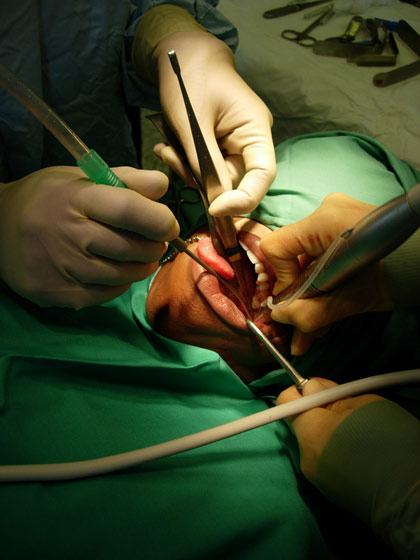
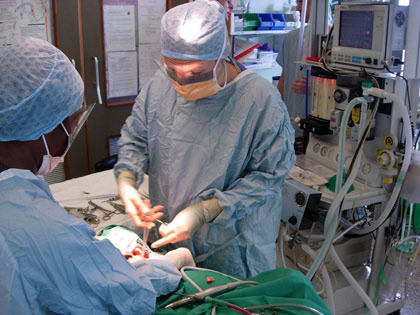
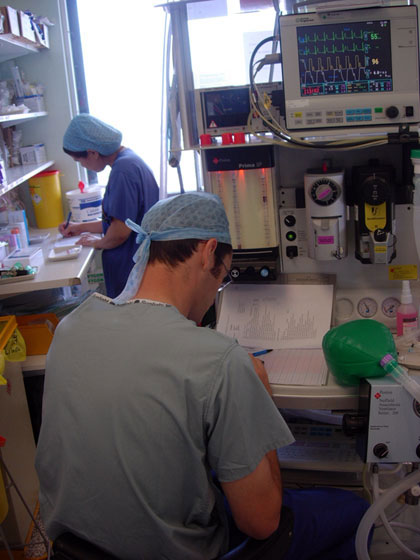
3. A surgeon and technician work together once the patient is anaesthetised.
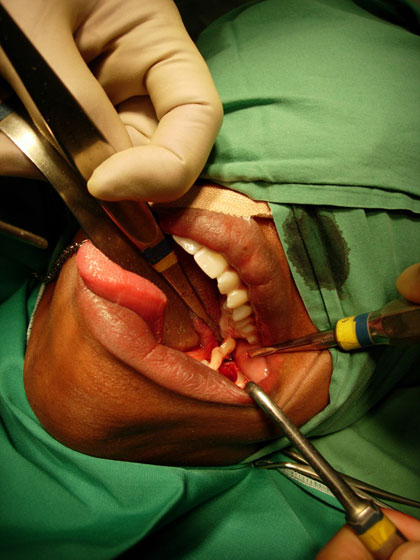
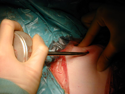
4. An insision on the gum exposes the tooth and allows it to be taken out.
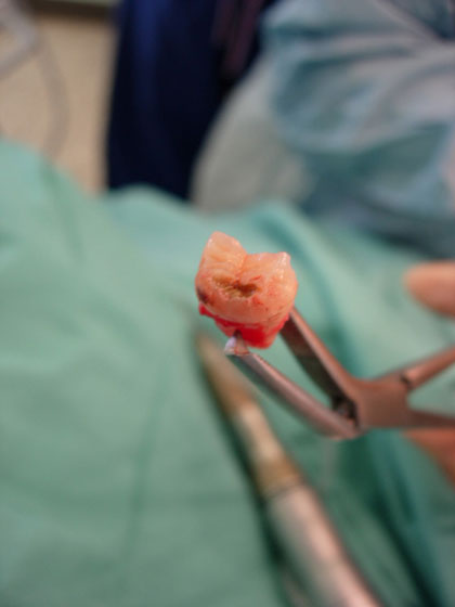
5. This tooth was too large to come out in one piece. It was drilled and split by a tool for removal.
6. Fluids are passed into the patients lungs to anaesthetise the patient. This process is monitored on the display behind the surgeon.
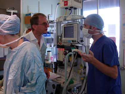
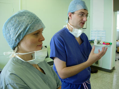
7. The anaesthetist amd surgeon work inependently. Here the anaesthetist describes his role to Tobie.
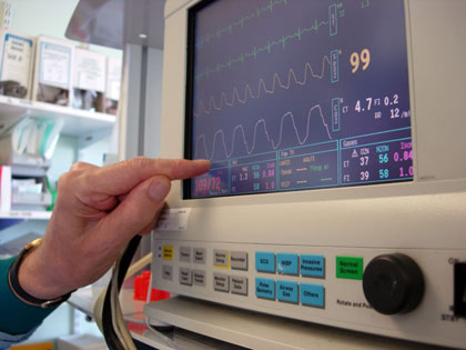
8. The four waves give an indication of the patients condition, the yellow waves shows the concentration of oxygen in the patients blood.
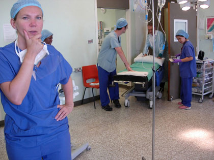
9. The surgeon reviews the work to be carried out on the next patient.
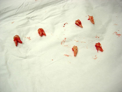
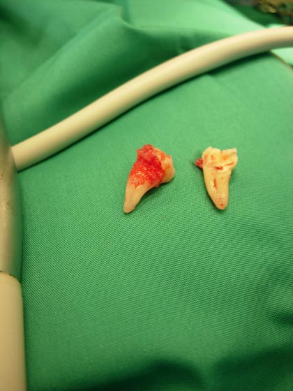
10. The four teeth from the first patient.
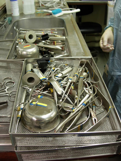
11. Surgical instruments are grouped by banded colours. Each group is used for one proceedure then sterilised.
12. Nikki and Ian discuss the operation with the surgeon as the first patient is woken up.
13. The theatre is prepared for the next patient.
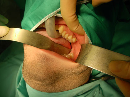
14. The problem tooth is on the lower jaw.
15. The large tube carries the anaesthetic, the transparent tube across the patients chest takes fluid out of his mouth.
16. A short break for the first patients anaesthetist.
17. Decay is clearly visible on the tooth.
18. The anaesthetist for the second patient records changes in the activity.

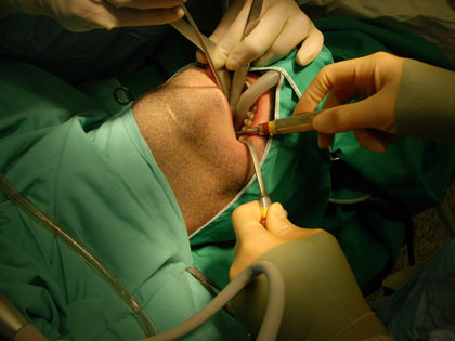
19. The surgeon and technician stitch the cavity left by the tooth.